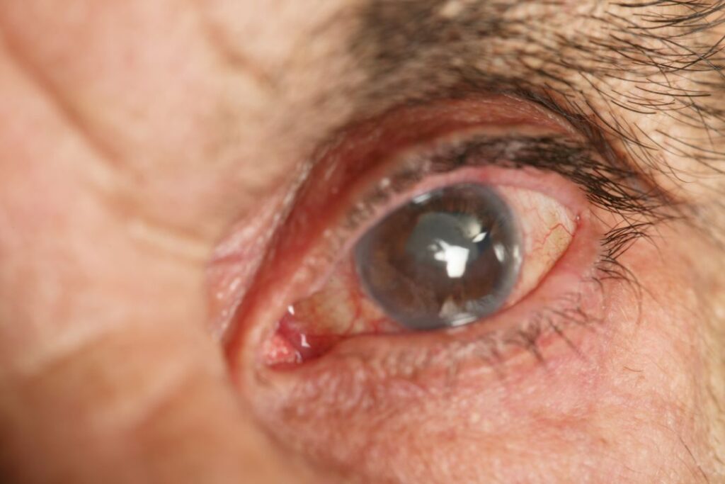
The retina is a vital component of our visual system, playing a crucial role in capturing and transmitting visual information to the brain. Understanding the structure and function of the retina is essential in identifying and treating common retinal conditions. This article will explore various aspects of the retina, common retinal conditions, their symptoms, diagnosis, and the available treatment options.
Understanding the Retina and Its Function
The retina is a fascinating and complex part of the eye that plays a crucial role in our vision. Let’s delve deeper into the anatomy and function of this remarkable tissue.
Anatomy of the Retina
In the context of cataract eye surgery, it’s essential to understand the anatomy of the retina. Positioned at the posterior part of the eye, the retina is a delicate and thin layer with distinct functions. At its outermost layer reside specialized cells known as photoreceptors, pivotal in capturing light and converting it into electrical signals.
Delving into the photoreceptor layer, we encounter two key cell types: rods and cones. Rods, highly sensitive to light, excel in low-light conditions such as nocturnal vision. On the flip side, cones, less light-sensitive, take the lead in color vision and visual acuity.
Descending beneath the photoreceptor layer, we navigate through critical layers involved in processing and transmitting visual information. These encompass bipolar cells, ganglion cells, as well as horizontal and amacrine cells. Each layer contributes to the intricate web of connections and interactions fundamental to the vision process.
In the realm of cataract surgery Sydney, comprehending the intricacies of retinal anatomy becomes particularly relevant for a holistic understanding of vision restoration.
Role of the Retina in Vision
The retina is a key player in the visual pathway, working in conjunction with other parts of the eye to enable us to see and understand our surroundings.
When light enters the eye, it first passes through the cornea and lens, which help to focus the light onto the retina. The photoreceptors in the retina then detect the light and convert it into electrical signals. This conversion process is essential for the brain to interpret and make sense of the visual information.
The rods and cones in the retina play different roles in vision. Rods, with their high sensitivity to light, are responsible for our ability to see in dimly lit environments. They provide us with black-and-white vision and help us navigate in low-light conditions. Cones, on the other hand, are responsible for our color vision and visual acuity. They allow us to perceive the vibrant hues and details of the world around us.
Once the photoreceptors capture the light and convert it into electrical signals, these signals are transmitted through the other layers of the retina. The bipolar cells and ganglion cells in the inner layers of the retina help to process and refine the visual information before it is sent to the brain.
The final destination for the visual signals is the optic nerve, which carries the information from the retina to the brain. The brain then processes these signals, integrating them with other sensory inputs, and forms a visual perception. This perception allows us to recognize faces, read text, appreciate art, and navigate our environment.
Understanding the intricate workings of the retina and its role in vision is crucial for appreciating the complexity of our visual system. It is a testament to the remarkable design and functionality of the human body.

Identifying Common Retinal Conditions
The human eye is a complex organ that allows us to see the world around us. Within the eye, the retina plays a crucial role in capturing light and sending visual information to the brain. However, there are several retinal conditions that can affect the normal functioning of this vital part of the eye. Let’s take a closer look at some of the most common retinal conditions:
Age-Related Macular Degeneration
Age-related macular degeneration (AMD) is a progressive condition that primarily affects older adults. It specifically targets the macula, a small area in the central part of the retina responsible for sharp and detailed vision. As AMD progresses, it can lead to a loss of central vision, making it difficult to perform everyday tasks such as reading or recognizing faces. Visit https://axonhealthcareinvestments.com/post-cataract-surgery-care-essential-tips-for-a-smooth-recovery-and-maintaining-optimal-eye-health/ to read about Post-Cataract Surgery Care: Essential Tips for Smooth Recovery and Maintaining Optimal Eye Health.
Early symptoms of AMD may include blurred or distorted vision, making it challenging to see fine details. Some individuals may also experience difficulty recognizing faces or reading small print. Another common symptom is the appearance of dark or empty spots in the central visual field, which can interfere with normal vision.
Diabetic Retinopathy
Diabetic retinopathy is a retinal condition that develops in individuals with diabetes, particularly those with poorly controlled blood sugar levels. Over time, high blood sugar levels can damage the blood vessels in the retina, leading to various changes that can affect vision.
One of the early signs of diabetic retinopathy is fluctuating vision, where visual acuity may vary throughout the day. Blurred vision is also a common symptom, as the damaged blood vessels can cause fluid leakage into the retina. Dark spots or floaters may appear in the visual field, and in severe cases, complete vision loss can occur.
Retinal Detachment
Retinal detachment is a serious condition that occurs when the retina detaches from the back of the eye. This detachment disrupts the normal flow of visual information from the retina to the brain and can result in permanent vision loss if not promptly treated.
There are several symptoms that may indicate retinal detachment. One of the most noticeable signs is the sudden appearance of floaters, which are tiny specks or cobweb-like shapes that seem to float across the visual field. Flashes of light may also be experienced, often described as seeing brief sparks or lightning-like streaks. Another significant symptom is the presence of a curtain-like shadow over a portion of the visual field, indicating that the detached retina is obstructing the normal visual pathway. Additionally, a significant decrease in vision may occur, making it difficult to see clearly.
Individuals who are nearsighted, have a history of eye injuries, or have undergone certain eye surgeries are at a higher risk of developing retinal detachment.
It is important to note that these are just a few examples of common retinal conditions. If you experience any changes in your vision or have concerns about your eye health, it is always best to consult with an eye care professional for a comprehensive evaluation and appropriate management.
Symptoms and Diagnosis of Retinal Conditions
Common Symptoms of Retinal Diseases
Although specific retinal conditions may have unique symptoms, there are some common signs to be aware of. These include changes in vision such as blurriness, distorted or wavy vision, dark or empty spots in the visual field, the sudden appearance of floaters or flashes of light, and a decrease in overall visual acuity. If you experience any of these symptoms, it is important to consult an eye care professional for a proper diagnosis.
When it comes to retinal diseases, understanding the symptoms is crucial for early detection and timely treatment. Blurriness in vision can occur due to retinal conditions such as macular degeneration or diabetic retinopathy. This blurriness may make it difficult to read or recognize faces. Distorted or wavy vision is often associated with conditions like retinal detachment or macular pucker, where the shape of objects may appear distorted or bent. Dark or empty spots in the visual field can be a sign of retinal holes or tears, while the sudden appearance of floaters or flashes of light may indicate a retinal detachment or vitreous detachment. Lastly, a decrease in overall visual acuity, or sharpness of vision, can be a symptom of various retinal diseases, including retinitis pigmentosa or retinal vein occlusion.
Diagnostic Procedures for Retinal Conditions
Eye care professionals utilize various diagnostic procedures to assess and diagnose retinal conditions. These procedures may include a comprehensive eye examination, visual acuity tests, dilated eye examinations, fundus photography, optical coherence tomography (OCT), fluorescein angiography, or electroretinography (ERG). These tests allow the ophthalmologist to evaluate the structure and function of the retina, aiding in accurate diagnosis and treatment planning.
A comprehensive eye examination is a crucial first step in diagnosing retinal conditions. During this examination, the eye care professional will evaluate various aspects of your eye health, including the retina. Visual acuity tests, such as the Snellen chart, measure your ability to see details at various distances. Dilated eye examinations involve the use of eye drops to widen the pupil, allowing for a better view of the retina. This procedure helps detect any abnormalities or signs of disease. Fundus photography is a non-invasive imaging technique that captures detailed images of the retina, providing valuable information about its structure and any potential abnormalities. Optical coherence tomography (OCT) is another imaging technique that uses light waves to create cross-sectional images of the retina, helping to diagnose conditions like macular edema or macular holes. Fluorescein angiography involves the injection of a dye into the bloodstream, which highlights the blood vessels in the retina. This test helps identify any abnormalities or blockages in the blood flow. Electroretinography (ERG) measures the electrical activity of the retina in response to light stimulation, aiding in the diagnosis of conditions like retinitis pigmentosa or cone dystrophy.
Treatment Options for Retinal Conditions
Medication and Drug Therapies
In some cases, medication and drug therapies can help manage retinal conditions. For instance, individuals with age-related macular degeneration may benefit from anti-vascular endothelial growth factor (anti-VEGF) injections, which can slow down the progression of the condition and potentially improve vision. Diabetic retinopathy may be managed with medications that help reduce swelling or control blood sugar levels.
Surgical Interventions
Surgical intervention is often necessary for retinal conditions such as retinal detachment or advanced cases of macular degeneration. Retinal detachment may require surgery to reattach the retina and prevent further vision loss. Advanced macular degeneration cases may benefit from procedures like vitrectomy, laser surgery, or retinal translocation. These surgeries aim to preserve or restore vision, and the choice of surgery depends on the severity and type of retinal condition.
Lifestyle Changes and Preventive Measures
In addition to medical and surgical interventions, certain lifestyle changes and preventive measures can help manage and reduce the risk of retinal conditions. These include maintaining a healthy diet rich in antioxidants and nutrients, regular exercise, managing blood sugar levels for individuals with diabetes, wearing protective eyewear when engaging in activities that pose a risk to the eyes, and avoiding smoking and excessive alcohol consumption. Click here to read about Perceived Barriers to Care and Attitudes about Vision and Eye Care: Focus Groups with Older African Americans and Eye Care Providers.
The Role of Regular Eye Examinations
Importance of Early Detection
Regular eye examinations play a vital role in the early detection and prevention of retinal conditions. Through comprehensive eye examinations, eye care professionals can assess the health of the retina, detect any early signs of retinal conditions, and intervene promptly. Early detection allows for timely treatment and management, which can prevent further vision loss and optimize visual outcomes.
Frequency of Eye Examinations
The frequency of eye examinations may vary depending on an individual’s age, medical history, and risk factors. Generally, adults should have a comprehensive eye examination at least once every two years, while individuals with certain risk factors or pre-existing eye conditions may require more frequent examinations. It is essential to consult an eye care professional to determine the appropriate frequency of eye examinations for your specific situation.
In conclusion, common retinal conditions can significantly impact an individual’s vision and quality of life. Understanding the structure and function of the retina, being aware of common retinal conditions, recognizing their symptoms, and seeking timely diagnosis and treatment are crucial for preventing vision loss. Regular eye examinations, adopting a healthy lifestyle, and following recommended preventive measures can aid in maintaining optimal retinal health. If you experience any changes in your vision or have concerns about your retina, it is crucial to consult with an eye care professional for expert evaluation and guidance.

